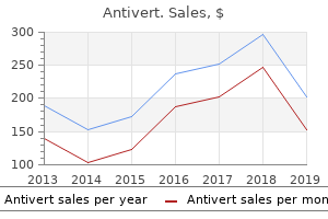
Antivert
"Purchase 25mg antivert mastercard, medicine 20th century".
By: X. Cobryn, M.B.A., M.D.
Associate Professor, Dell Medical School at The University of Texas at Austin
By far the most common cause of acute renal failure is hypoperfusion of the kidney symptoms of dehydration generic antivert 25mg otc, referred to as pre-renal acute renal failure daughter medicine purchase antivert uk. This is usually seen with hypovolaemia secondary to conditions such as acute blood loss, vomiting, diarrhoea and burns. It may also occur in normovolaemic patients with sepsis, cardiac failure or renal artery stenosis. Investigation demonstrates high serum urea, creatinine and potassium concentrations, and a poor urine output. If the kidneys are hypoperfused for a significant amount of time, autoregulatory mechanisms within the renal vasculature fail, causing tubular cell damage and death. During recovery, there is an oliguric phase (when both glomerular filtration and tubular function are compromised), followed by a diuretic phase (when glomerular filtration recovers before tubular function is regained). This condition is classically seen in childhood following gastroenteritis caused by Escherichia coli O157, which produces nephrotoxic verotoxins. The haemolytic anaemia and thrombocytopenia are due to erythrocyte and platelet destruction within fibrin meshes that are deposited on affected endothelial membranes. Occasionally, renal function is permanently compromised, necessitating long-term renal replacement therapy and renal transplantation. Urinary tract obstruction may be caused by prostatic hypertrophy, urinary tract calculi, renal tract malignancy, constipation or retroperitoneal fibrosis. In this case, the patient presented with urinary retention secondary to prostatic disease. The obstruction causes urine to accumulate under high pressure in the renal pelvis, resulting in a deterioration in renal function. In cases of urinary retention secondary to prostate pathology, a urinary catheter should be inserted to bypass the obstruction until a more definitive treatment can be organized. The arterial stenosis results in renal hypoperfusion, which in turn stimulates the secretion of renin from the juxtaglomerular cells of the kidney. The patient often has significant diarrhoea, which can lead to rapid dehydration, electrolyte imbalance and death. Much effort is being put into in the prevention of clostridial infection by promoting the correct use of broad-spectrum antibiotics and educating hospital staff and visitors regarding the importance of hand washing. It can usually be managed conservatively, but occasionally requires a course of erythromycin. Infection is usually due to consuming rewarmed rice that has been contaminated with spores. The patient may have a prodrome of upper respiratory tract illness followed by development of diarrhoea and vomiting. The illness is self-limiting and usually requires no treatment other than supportive measures.
Cajal (1989) reasoned that the central and the peripheral expansions of these unipolar cells could still be considered as "functioning in the same manner as the two expansions of the treatment 3rd degree av block order discount antivert. He studied in Louvain (Leuven) medications borderline personality disorder discount 25 mg antivert amex, which numbers Vesalius among its illustrious alumni. While there he began his training in microscopical anatomy under Jean-Baptiste Carnoy, a well-known anatomist, author of Biologie Cellulaire (Cellular Biology) and editor of the journal La Cellule. Van Gehuchten took further training in Berlin and the Law of Dynamic Polarization 203 Frankfort-am-Main in the 1880s, and then returned to Louvain in 1887, at the age of 26, to a chair of anatomy. Van Gehuchten published two long papers in La Cellule supporting this idea, on invertebrate muscle (1886) and vertebrate muscle (1888). I was corresponding with Cajal in connection with papers which each of us had published upon the finer structure of the muscle cell. One day he wrote to me announcing that he was abandoning his investigations upon muscles in order to devote himself to the nerve centres, basing his decision upon the fact that he had obtained remarkable results from the application to embryos of one of the formulae of the method of Golgi, which had been discovered in 1875. Van Gehuchten immediately abandoned his previous work to follow Cajal in applying the Golgi method to the nervous system. When the two met at the Berlin Congress in late 1889, van Gehuchten had not yet published his first paper on the nervous system, but Cajal regarded him as the "young and already brilliant professor of anatomy from the University of Louvain" (he would have been 28 by then), and one whose approval he sought and valued. But all nerve cells with all these prolongations form an independent element, a whole autonomy, a kind of nerve unity. This independence of the nerve elements from each other has moreover been proved embryologically by the beautiful research of His. This learned man has shown that one nerve cell with all these prolongations comes solely from the transformation of a neuroblast. When one researches in classical books which are the component elements of the nervous tissue, one ascertains that everywhere nerve cells and nerve fibers are considered as two absolutely distinct and completely independent structures. This is so true that, for many years, authors have been forced to search for the method of union and the method of action of the nerve fibers and the cell fibers. From new notions on the structure of the nerve centers, it clearly results that the nerve fibers and the nerve cells are not distinct elements: the nerve fiber considered by itself and in its essential part, the axis-cylinder, is not a nerve element, no more than the protoplasmic prolongations of the nerve cells: it is only a cylindraxil prolongation. The nerve cell in itself is not a nerve element either, one cannot separate it from either its protoplasmic prolongations, if these exist (this qualification is necessary because the nervous elements of the spinal ganglia are deprived of protoplasmic prolongations [dendrites]), or from its cylindraxil prolongation. The sole nerve element is the nerve cell with all its prolongations [italics added]. The nerve elements, thus composed, vary infinitely in their form, volume, and disposition and richness of the protoplasmic prolongations [dendrites]; only one of their characteristics seems constant and permits one to distinguish a nerve element from all other elements: it is the existence of a cylindraxil prolongation [axon]. This cylindraxil prolongation has particular characteristics which are rather difficult to describe, but which however permit one to recognize it at first sight, whatever may be the number of protoplasmic prolongations from the nerve cell. It springs directly from the cell body or arises, sometimes at a very great the Law of Dynamic Polarization 205 distance, from one or another of the protoplasmic prolongations. Before we carried out our own research with the Golgi method, we often asked ourselves, while examining the stained nerve cells in the work of Golgi and his students, why a given prolongation, colored in red by the author, was the cylindraxil prolongation, since it seemed not to differ by any of its characterizations from the neighboring prolongations, and since quite often one or the other of these, longer or more voluminous, deceived us at first. This same observation we have heard detailed many times by several of our colleagues. The cylindraxil prolongation, in our opinion, is distinguished from protoplasmic prolongations above all by the sharpness of its contours and by its regular trajectory. If it emits a collateral branch, or if it bifurcates, one always finds at the point of division a small triangular thickening with regular contours like the axis cylinder itself. At any rate, the only way to familiarize oneself with the characteristics of the cylindraxil prolongation, in order to recognize it without any difficulty amidst the protoplasmic prolongations of a nerve cell and compare it at the same time with the drawings of other authors, is to make some attempts oneself with the Golgi method. A few good reductions [stains] will suffice to convince the most skeptical of the completely specific characteristics of the cylindraxil prolongation. It behaves everywhere in the same way, ending freely by one or several terminal branches. There are nerve cells whose cylindraxil prolongation is short and ends very near the cell body: there are others whose cylindraxil prolongation extends over a considerable length: such, for example, is the cyilndraxil prolongation of the radicular cell [motor neuron], which extends from the grey matter of the spinal cord to the peripheral muscle; but this morphological distinction is not clear enough to see, with Golgi, evidence for a physiological distinction; these are only two extreme forms between which one finds a whole series of intermediary forms. All the central nervous system thus reduces itself, in the final analysis, to a superposition of independent nerve elements. Among the nerve elements with long axis cylinder [axons], one can easily distinguish two types at first sight. One type has its cell body in the superior parts of the cerebro-spinal axis, and its cylindraxil prolongation [axon] descends to end freely lower down.
If the Mediterranean lay in the penumbra of European academic activity xanax medications for anxiety generic antivert 25mg visa, how much further away was America treatment yeast overgrowth antivert 25 mg with mastercard, and how much more keenly felt the distance. In America, we frequently find recent graduates who tell us that they would follow an academic career if their future were assured as far as salary is concerned. Little do they realize that this attitude of mind alone should exclude such a career. Mall himself was one of the early students in the United States who made the trip to Europe, to study under His; his remarks indicate how important were continental institutions as the seedbed for research training and as an ideal for academic enlightenment. They served as the model for the development of academic research laboratories throughout the world well into the twentieth century; indeed, modern physics arose from just such a network of outstanding researchers and peripatetic students in central Europe during the 1920s. Only toward the end of the nineteenth century did the balance in the biological and medical sciences begin to shift to Great Britain and, following the devastation of the First World War, to the United States. Describing Nerve Cell Development In 1857, His, at the age of 26, won appointment to the chair of anatomy at Basel. Again, he was blessed with freedom to pursue his interests, and he knew how to use it, launching a comprehensive program of study that laid the foundations of the cellular basis of embryology. Central to this effort was the concept of the three germ layers as providing the origins for the different tissues and organs of the body. He devised a microtome for cutting serial sections of high quality through an entire embryo. In 1872, he was called to the chair of anatomy at Leipzig, where he pursued his studies even more vigorously, aided by improved facilities and technical support. Soon he turned his attention to human development, and between 1880 and 1888 published another monumental three-volume work, Anatomie menschlicher Embryonen (Anatomy of the Human Embryo). During this period, at the peak of his investigative career, His focused his attention on the development of nerve cells in the spinal cord. He was well aware of the A Neuron Theory Begins to Take Form: His, Forel, Nansen 103 difficulties of visualizing nerve cell processes in the adult, and of the controversies surrounding the theories of Gerlach and the others who believed in anastomoses and continuity of the processes. He realized that analysis of the cells early in development, before they formed their processes and during the formation of the processes, would provide a way of testing the theory. In an early section of the paper, he describes the outgrowth of processes from the nerve cell: the nerve fibers extending from the cells start in the vicinity of a nuclear pole with a conically broadened base and show clear fibrillar stripes at their origin. The cell body extends only a little over the nucleus and where it is possible to see it somewhat freely, it appears to be trimmed with several blunt points. Accordingly, the formation of the branched extensions [dendrites] occurs considerably later than that of the axis cylinder extension [axon]. Here is an unequivocal description of what is seen, not a speculation on what can be imagined. The early outgrowth of the axis cylinder (axon) followed by later outgrowth of "branched extensions" (dendrites) is a rule of nerve cell development that has generally been confirmed by modern studies. One feels in reading this paper that one is finally on the road to modern publication of experimental results and conclusions. Here a controversy centered on whether the fibers of this root arise from cells within the spinal cord or from the cells within the dorsal root ganglia (see Freud, Chapter 7). His again came forward with observations and a conclusion: In an earlier communication, I presented the reasons which then favored growth of the sensible roots from the ganglia into the spinal cord. These were: 1) the trophic dependence of the root fibers on the ganglion as shown in experiments by Waller and von Bernard; 2) the early appearance of a fibrillation inside the ganglion not connected with the spinal cord; 3) the beginning shape of the upper root part becoming pointed towards the spinal cord; 4) the fact that the sensible fibers in the spinal cord have another fiber direction beyond these. A further problem with the dorsal root ganglion cell was the fact that in the adult the cell appears unipolar, that is, the cell body gives rise to a single smooth process, which, after a short distance, divides into two branches, thus having a T-shaped form. As discussed in Chapter 7, this branching pattern was initially puzzling to anatomists, who, first of all, could not understand how it could be reconciled with the pattern of single axis cylinder (axon) and multiple protoplasmic extensions (dendrites) that seemed to be the rule elsewhere in nerve cells, and second, were hard put to give it a functional interpretation.
That is exactly what happened in the first half of the 20th century; interest turned to other types of cell stains and other kinds of issues symptoms gluten intolerance buy antivert on line. The Nissl stains treatment hiccups order discount antivert on line, for example, which reveal the nuclei and parts of the cytoplasm of all neurons, became widely used to classify the cerebral cortex into numerous local areas on the basis of fine distinctions in the staining pattern of different cell layers. Without it, the whole thrust of analysis of questions surrounding the neuron in the classical period faded. What was needed, of course, in order to make any further progress on the unresolved issues, was the introduction of new methods. However, by the early 1950s anatomists had acquired the electron microscope for high-resolution analysis of cell structure, while physiologists had developed the microelectrode for intracellular 279 analysis of the membrane mechanisms of nerve impulses and synaptic potentials, as well as for extracellular recordings of single cell activity for analysis of information processing in neural circuits. These tremendous changes recall the great advances in biology that had been made around the 1880s and, before that, around the 1840s. To discuss the introduction of these modern methods would be to chronicle the rise of modern neuroscience, which is beyond our scope. Our aim in this chapter will therefore be to focus narrowly and briefly on those advances that had specific relevance to the neuron doctrine. We will organize the discussion around the four tenets originally put forward by Waldeyer: that the neuron is the anatomical, physiological, genetic, and metabolic unit of the nervous system. To what extent were these tenets supported, modified, or disproved by the early work of the modern era Much of the theory stood the test, and has served well as the basis for the remarkable advances that have occurred in recent decades. However, some surprises occurred that had not been anticipated by the classical concepts. We will summarize the attempts that were made to deal with these, and finally indicate some future directions in which revisions are likely to be needed. What was needed was an entirely new type of microscope with much higher resolving power. Whereas the light microscope forms images using light waves, which limits the resolving power in most circumstances to the wave length of light (slightly less than one micron-one thousandth of a millimeter), the electron microscope, as its name implies, forms images by using beams of electrons, whose resolving power is about 10,000 times greater. The application to animal tissue encountered great difficulties in obtaining adequate fixation and staining of the tissue so that it would give clear images while withstanding the high intensity of the electron beams. These difficulties were most extreme with nervous tissue, but gradually the images furnished the long-awaited answers. The Synapse the most critical point of attack in assessing the question of continuity or contact between neurons was the synapse. One strategy was to study the neuromuscular junction, where the difference between the nerve terminal and the muscle cell is marked. At central synapses, an important clue was the high density of mitochondria in most axon terminals. At these sites, there is a presumed physiological sequence of activity, such that the axon terminal is the presynaptic process, and the soma or dendrite that it contacts is the postsynaptic process. These findings were immediately interpreted as providing a definitive resolution of the old question of neuron versus reticulum. This same verdict was reached by another pioneer, Eduardo de Robertis (1959): the observation of a neat delimitation of both the pre- and postsynaptic cytoplasm confirms and extends to a submicroscopic level the concept of the individuality of the nerve element which is implicit in the neuron doctrine of Cajal. The reticularist hypothesis, which still has its followers, cannot be maintained, even in those regions of the central nervous system called neuropiles, where most of the elements are of submicroscopic dimensions. The reticular appearance is the result of technical artifacts, plus the limited resolving power of the optical microscope to detect those structures and their boundaries. These facts indicate that for an exact interpretation all structures below 1 to 0. Modern Revisions of the Neuron Doctrine 281 Note that, in both these cases, the credit for the neuron doctrine is given to Cajal; the citations, in fact, were to his last writings on the subject in 1933. These early electron-microscopic studies of the synapse constitute one of the great discoveries in neuroscience.


