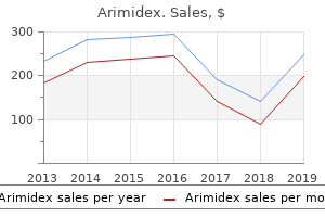
Arimidex
"Purchase arimidex master card, women's health clinic evergreen park".
By: H. Owen, M.A., M.D., M.P.H.
Clinical Director, Sanford School of Medicine of the University of South Dakota
Less frequently menstruation vs miscarriage discount generic arimidex canada, a papillary adenocarcinoma arises from a polyp or pedunculated adenoma and invades the gastric wall through the stalk breast cancer uggs arimidex 1mg discount. Some adenocarcinomas assume on their surface a polypoid or fungating appearance, with necrotic and ulcerating foci. On the cut section, this "vegetative" type of carcinoma, as it has been called, presents a yellowish, solid mass in a gray fibrillar stroma. The histologic architecture of the adenocarcinoma may sometimes exhibit the typical columnar cell arrangement, with formation of glandular spaces, but it is usually more complicated and varies considerably. Atypical tubular glands may replace the normal mucosal pattern, penetrating into the muscularis mucosae or spreading from the submucosa as far as the serosal coat. At times, the tumor consists only of closely grouped alveoli with cylindrical and cuboidal cells and hyperchromatic nuclei. The cells lining these alveoli may, in some cases, contain substantial amounts of mucus, and, occasionally, the entire tumor may be replaced by gelatinous or slimy colloid material, in which only a few embedded cancer cells may be found. In such instances, the displaced nuclei and overextended, ruptured, or disintegrated cells in this mucinous matrix may create a most complex histologic picture. This rare type of stomach cancer begins in the lining of the stomach and spreads to the muscles of the stomach wall. This causes the wall to become thick, hard, and rubbery, which leads to trouble in digesting food. Another cause of linitis plastica is metastatic infiltration of the stomach, particularly by breast or lung carcinoma. Linitis plastica produces a diffuse thickening of all layers and involves a large part of the gastric wall (sometimes, the entire wall), which becomes contracted and rigid. The scirrhous malignant lesion usually begins in the pyloric canal and may, in some cases, remain limited to this region, where it may soon cause signs of obstruction, because the profuse growth of its fibrotic components markedly reduces the lumen. The same phenomenon takes place over the whole gastric cavity, when the scirrhous growth has expanded extensively over the entire lining. The mucosal folds become immobile and inflexible, while simultaneously, as a result of the abundant formation of fibrous tissue, the whole organ shrinks, assuming a shape that has been described as the leather bottle stomach. Histologically, nests of epithelial cells are scattered in dense fibrous tissue, which leaves nothing of the normal gastric structures. The number of recognizable malignant cells is gradually reduced, and, in the advanced stages, it is difficult to demonstrate their presence except by the most painstaking microscopic study. In some cases the fibrotic reaction has gone so far as to Malignant infiltration limited to pylorus make recognition of the original nature of the process practically impossible. In view of such proliferation of connective tissue, it is not surprising that the primary cause was formerly considered to be a chronic reactive inflammatory process and received, accordingly, the designation linitis plastica. The roentgenographic appearance of linitis plastica, or scirrhous carcinoma, varies, of course, depending upon the extent to which the gastric wall has become involved. If limited to the pyloric region, a localized area of narrowing, distinct irregularities of the contour, and the disappearance of the normal mucosal markings leave no doubt as to the diagnosis. If, however, the neoplasm has spread over a larger segment or, as happens not infrequently, over the entire inner aspect of the stomach, the cavity of the stomach presents itself as a narrow tube with no mucous membrane pattern visible. The contour in such cases may be erratically distorted, and the barium meal rushes through the organ because of the rigidity of the pylorus, which, under these circumstances, is permanently opened. Because the obstruction in advanced linitis plastica is located at the cardia, it is the esophagus that eventually becomes dilated. With x-ray findings as clear as those described above, the diagnosis of scirrhous carcinoma presents no difficulties, and laboratory data, such as achlorhydria, hypochromic or hyperchromic macrocytic anemia, or occult blood resulting from the destruction of the glands or from erosions, provide little more than mere additional supporting or confirming information. Upper endoscopy, at times difficult to perform because of the rigidity and lack of air in the stomach, may help establish the diagnosis, although the endoscopic picture of an infiltrating carcinoma may now and then resemble that of a lymphoma or hypertrophic gastritis, necessitating a biopsy for differentiation. The unfortunate feature of the situation, however, is that these characteristic x-ray pictures are seen only in a late stage of the disease when the presence of lymph node metastases can be expected. Symptoms develop rather insidiously, and patients come for medical care at a time when total gastrectomy, the only sensible treatment for this condition, can scarcely be more than palliative. The prognosis may become more favorable for the infiltrating type of carcinoma, as for other types of cancer of the stomach, when the methods for early recognition improve and when institutions such as cancer prevention clinics are more widely used. Necroses and loss of substance on the surface of a diffuse, infiltrating, scirrhous carcinoma are relatively rare and only superficial, whereas the funguslike, proliferating, and more circumscribed (but still broadly infiltrating) neoplasms tend often to become deeply ulcerated by the sloughing of substantial parts of their central segments, probably because their blood supply cannot keep pace with their rapid growth. In such cases, especially with the early superficially spreading type, it may be extremely difficult, if not impossible, to separate the ulcerating carcinoma diagnostically from a benign chronic, callous, and penetrating peptic ulcer.
The entire joint can be visualized by sweeping the scope from the radial to the ulnar side and rotating the scope as necessary breast cancer forum order genuine arimidex on-line. The volar central radiocarpal portal is established underneath the lunate through the interval between the ulnocarpal ligaments and the short radioulnar ligament breast cancer network of strength cheap arimidex 1mg with mastercard. With the arthroscope in the 6R portal, a 22-gauge needle is introduced just under the lunate. The volar capsule is pierced with tenotomy scissors, followed by insertion of the cannula into the radiocarpal joint. The joint is visualized similarly by sweeping the scope from the radial to the ulnar side. The forearm is held in supination to relax the dorsal capsule, to move the ulnar head volarly, and to lift the central disc distally from the head of the ulna. The joint space is identified by first inserting a 22-gauge needle horizontally at the neck of the distal ulna. The joint is infiltrated with saline, and the capsule is spread with tenotomy scissors through a small incision. Because of the dorsal entry of the arthroscope, the course of the dorsal radioulnar ligament is not visible until its attachment into the fovea is encountered. This is especially useful for arthroscopic assisted fixation of distal radius fractures. Multiple examples of dry arthroscopy for the wrist and small joints can be seen through this book. It is still necessary to use fluid when a thermal probe is used, to minimize the risk of heat necrosis of the articular cartilage. Summary Advances in wrist arthroscopy continue to expand the indications and treatment options for myriad wrist disorders. A systematic approach and a thorough understanding of the topographical and internal anatomy of the wrist are integral to minimizing complications while maximizing the chances of a successful outcome. Arthroscopic resection of arthrosis of the proximal hamate: A clinical and biomechanical study. Intraarticular fractures of the distal aspect of the radius: Arthroscopically assisted reduction compared with open reduction and internal fixation. The overlap pattern of the lateral antebrachial cutaneous nerve and the superficial branch of the radial nerve. Rehabilitation the postoperative rehabilitation is dictated by the treatment of the specific pathology. Following an arthroscopic debridement alone, wrist range of motion exercises are instituted within 3 to 5 days postoperatively. The cutaneous innervation of the palm: An anatomic study of the ulnar and median nerves. Anatomy of the palmar cutaneous branch of the median nerve: Clinical significance. Vascular and neural anatomy of the thenar area of the hand: Its surgical applications. Anatomical reduction of intra-articular fractures of the distal radius: An arthroscopically-assisted approach. Arthroscopic assessment of the volar region of the scapholunate interosseous ligament through a volar portal. Isolated tears of the triangular fibrocartilage: Management by early arthroscopic repair. Visualisation of the pisotriquetral joint through standard portals for arthroscopy of the wrist: a clinical and anatomical study. Berger independently developed his technique for arthroscopic evaluation of the first carpometacarpal joint, which he first presented as an instructional course in 1995. He named the volar radial portal the 1-R portal and the dorsal ulnar portal the 1-U portal (Video 2-1). The terms radial and ulnar referred to the thumb when its nail is parallel to the fingernails with the thumb supinated and radially abducted. Both portals are along the radial border of the thumb, which makes it difficult to assess the lateral side of the joint.
The volar medial fragment is reduced under direct observation by pinning it back to the shaft and the radial styloid fragment menstruation moon cycle discount arimidex master card. In this event womens health questions free arimidex 1 mg online, one or more of the distal screws should be placed in a nonlocking fashion to help compress the fragments. A Freer elevator is inserted dorsally through the 3,4 portal and introduced into the fracture line to disengage the dorsal lunate facet fragment. A large hook probe is introduced obliquely through the fracture line and under the volar cortex of the volar lunate facet fragment, which is then tilted, disimpacted, and reduced. In contrast to the management of dorsal lunate facet fragments, in this latter technique, reduction of the fragments is performed first and then the radial styloid is reduced. If the radial styloid is first reduced to the metaphysis, this will not allow further subsequent reduction of the volar lunate facet. Next the fracture is reduced and a volar locking plate is provisionally applied with K-wires. Many of the available volar locking plates have K-wire holes in the shaft and distal row for this purpose. The K-wires also provide a guide for the subsequent trajectory of the distal row screws. The hand is suspended in a traction tower and the fracture site reduction is observed under dry arthroscopy with the scope in the 6R portal and a full radius resector in the 3,4 portal. A fine-tuning of the articular reduction is then performed by backing out the distal K-wires, manipulating the fracture fragments with joysticks and an elevator, and then reinserting the K-wires. At this point the distal locking screws are inserted to support the articular fragments. This approach can also be applied to fine tuning of dorsally located intra-articular distal radius fractures (Video 11-6). This reference fragment was then reduced and stabilized with locking pegs or screws to an extra-long volar plate under fluoroscopic guidance. Once the fragment reduction and fixation were achieved, additional diaphyseal screws were inserted into the stem of the plate proximal to the comminution to secure the plate to the diaphysis. This provided a rigid construct and avoided migration during the arthroscopic portion of the operation. They then placed the hand in a traction tower and reduced the rest of the fragments to the reference fragment using dry arthroscopy. By securing the reference fragment before addressing the metaphyseal comminution, a stable platform was created. Once the articular reduction was complete, the comminuted metaphysis was addressed and secured to the plate. They reported good results in 4 patients, 2 requiring free flap coverage, and 3 requiring bone graft. Compared with the contralateral wrist, range of motion was 79% and grip strength was 90%. Eight patients had excellent results, 11 had good results, 7 had fair results, and 2 had poor results. Twenty-three patients were able to return to their previous activities level or occupation without any restriction. They concluded that an arthroscopic-assisted reduction combined with volar plating or external fixation is one of the useful options for the treatment of a displaced intraarticular fracture of the distal radius in active elderly patients. They concluded that an arthroscopic reduction of intraarticular fragments is superior to reduction under fluoroscopy, and it also permits the detection of associated intraarticular soft tissue lesions.

The one (network 10) included the middle and inferior temporal gyri and the middle temporal visual association area at the temporo-occipital junction menopause hormone levels discount 1 mg arimidex amex. This network corresponded to one activated in tasks involving the viewing of complex menstruation bible buy genuine arimidex line, often emotional, stimuli but also observation of actions, overt picture naming, and visual tracking of moving objects, mental rotation, and the discrimination of locations in space. Resting networks extracted and degree of concordance among studies expressed as the number of studies in which a given network was found. Network 11 corresponded to the one activated in the context of higher level visual processing tasks, such as those involving orthography and covert reading, as well as Braille reading tasks, whereas network 12 corresponded to tasks involving simple visual stimuli such as flashing checkerboards. The first consisted of the areas located in the calcarine sulcus bilaterally and of medial structures such as the lingual gyrus, the inferior part of the precuneus and the lateral geniculate nucleus of the thalamus. The second consisted of the lateral visual cortical areas, including the occipital pole extending laterally toward the occipitotemporal junction and the superior parietal regions of the cortex. It appears therefore that if there is a discrepancy among these findings, it consists only in the extent of the visual network reported across the 10 studies. The same network, Laird and associates found, is activated in the context of theory of mind and social cognition tasks and to a lesser degree in fixation, episodic recall, imagined scenes, and delay discounting tasks. He and his associates derived a similar network but one that also included the middle temporal gyrus; Yeo and associates (2011) derived one that encompassed, in addition, left temporal lobe areas; Damoiseaux and associates (2006) one that included both the anterior and the posterior cingulate gyrus, the inferior temporal and the superior parietal region but not the temporoparietal junction; and Allen and associates (2011) one that also included the middle frontal gyrus, bilaterally and the right inferior frontal gurus. Almost as reliable appear to be the "frontal," the "somatosensory-motor," and the "auditory" networks that were replicated in seven of the 10 studies reviewed here. This wide range of network functions was elicited across all possible stimulus modalities representing heterogeneous functions ranging, according to the authors, from executive to affective including basic sensory (interoceptive) ones. Three somatosensory-motor subnetworks were identified by Laird and associates (2011) that included the ventral precentral gyri, the central sulci, and the postcentral gyri, but also the superior and inferior cerebellum, the dorsal precentral gyri, the central sulci, the postcentral gyri, and the superior and inferior cerebellum. According to the authors, the somatosensorymotor network is activated in tasks involving action and somethesis corresponding to hand movements such as finger tapping, grasping, pointing, and electrical and vibrotactile stimulation, but also to speech tasks, such as overt reading or recitation, chewing or swallowing, and flexion/extension of the tongue. Similarly, DeLuca and associates (2006) derived a network that included, besides the pre- and postcentral gyri midline regions, the thalamus and the hippocampus. Yeo and associates (2011) derived one that included the supplementary motor, the motor, and somatosensory cortex down to the Sylvian fissure but the superior temporal region of the right hemisphere as well. It included the primary auditory cortices and was found to be activated in auditory tasks including tone and pitch discrimination, music, phonological discrimination, and oddball discrimination. Allen and his group (2011) derived subnetworks that fell, again, within the broader region as defined by the other studies. Considerable concordance among studies is found in the derivation of a family of networks that are sometimes called attentional and other times executive and involve frontal and parietal regions. This network is activated in visuospatial processing and reasoning tasks such as the Wisconsin Card Sorting Test, saccades, antisaccades, mental rotation, and counting or calculation. Another variant of this "attentional" network was derived by Yeo and his associates (2011) consisting of three subnetworks, the first involving superior prefrontal and superior parietal regions, the second inferior prefrontal and inferior parietal regions extending medially, and the third including the posterior part of the middle and inferior temporal cortex and, medially, the cingulate. A fourth variant, derived by DeLuca and his associates (2006), was composed of two distinct subnetworks, the one involving the lateral prefrontal and the dorsal parietal cortex and the other the inferior occipital, parietal, temporal, and inferior prefrontal cortices. It appears that there is considerable grounds for concluding that there might be two types of frontalparietal networks, the one referred to as "ventral" and the other as "dorsal" attention systems as those derived by Fox et al. On the other hand, one could argue that the frontal parietal network variants found in these studies may be identified with executive functions, given the ambiguity of the terms "attention" and "executive" functions as they are used in the neuroimaging literature. Only half of the studies reviewed here have derived variants of a left lateralized network. Variants of the same network were derived by Smith and associates (2009); by Beckmann and associates (2005), although in that study the network included the intraparietal region bilaterally; by Damoiseaux and his group (2006) that included the middle frontal and orbital, the superior parietal, the middle temporal gyrus, and the posterior cingulate of the left hemisphere; and by Allen and his associates (2011), which featured the left inferior middle frontal and the angular gyri but also the right superior parietal lobule. These areas are activated, according to the authors, by tasks involving reasoning, attention, inhibition, and memory, such as the n-back, delay discounting, and divided auditory attention tasks. A second, similar network but one including the insula was found by Smith and his group (2009); a third, including part of the right lateral occipital cortex, the right inferior parietal cortex, the right middle and superior frontal gyri, and both the right and the left intraparietal sulcus was derived by Beckmann and his associates (2005) who identified it with the "dorsal visual stream"; and a fourth, by Damoiseaux and his associates (2006) featuring predominantly right-hemisphere regions such as the middle frontal and orbital, the superior parietal cortex, the middle temporal, and the posterior cingulate gyrus, identifying them as part of the "function of memory. This network was found to be activated by tasks involving discrimination of emotional faces and pictures, particularly those that elicited fear, happiness, and humor, but also tasks involving "air-hunger," olfactory, and gustatory responses. Yeo and his group (2011) also identified a similar variant involving the medial aspect of the temporal tips and parts of the orbitofrontal cortex, as did Salvador and his associates (2005). The Default Mode and Other Resting State Net works 205 group, on the other hand, derived a more extensive network that included the left and the right parahippocampal gyrus, hippocampus, amygdala, the temporal pole, the olfactory cortex, the thalamus, the caudate, putamen, and the pallidum. Replicated in only two studies (Demoiseaux and his group [2006] and Laird and her group [2011]) are networks involving only the parietal regions; only the cerebellum (Laird and associates [2011] and Smith and associates [2009]), and only the basal ganglia (Laird and associates [2011] and Salvador and associates [2005]).


